Welcome to Ovarian Aging!
Today I will discuss what happens when ovaries age and how we can use this information to inform therapeutics for patients. For a fun visual learning experience I recommend watching my 4 minute video above.
After watching/reading you will learn about:
3 Pillars of Ovarian Aging
We’ve learned from my previous posts that ovarian aging affects reproduction, hormone production and overall health. But what does ovarian aging actually look like in the ovary? Here I will talk about the 3 pillars of ovarian aging.
Pillar 1: Age related decline in follicle quantity and quality
As the ovaries age, so do the oocytes (or eggs) inside follicles. Eggs have special machinery inside them that is used during cell division. This machinery ensures proper chromosome separation so each cell maintains the correct number of chromosomes as it divides. The machinery is built into the eggs when individuals with ovaries are born and does not get updated, so the older the eggs are the older the machinery is. Therefore ovarian aging is defined by an overall decline in follicle quantity and quality.
Pillar 2: Chronic Inflammation
The environment follicles are sitting in within the ovary starts to change with age as well. The ovarian microenvironment becomes increasingly inflammatory. While normal inflammation is a protective immune response, chronic or long lasting inflammation leads to damage. Over time, there is an increase in pro-inflammatory molecules in the ovary leading to a state of chronic inflammation.
Pillar 3: Ovarian Fibrosis
Additionally, the ovarian microenvironment becomes fibrotic with age. In the simplest terms, fibrosis is an increase in tissue stiffness. Aging disrupts the network of tissue in the ovary which becomes stiffer and leads to many problems including a disrupted ability to ovulate.
✨Repro Relevance✨
So why might this matter to you or someone you know?
We can use the research showing increased fibrosis and inflammation in ovaries with age to come up with potential therapeutics that may extend reproductive longevity. Here are two examples:
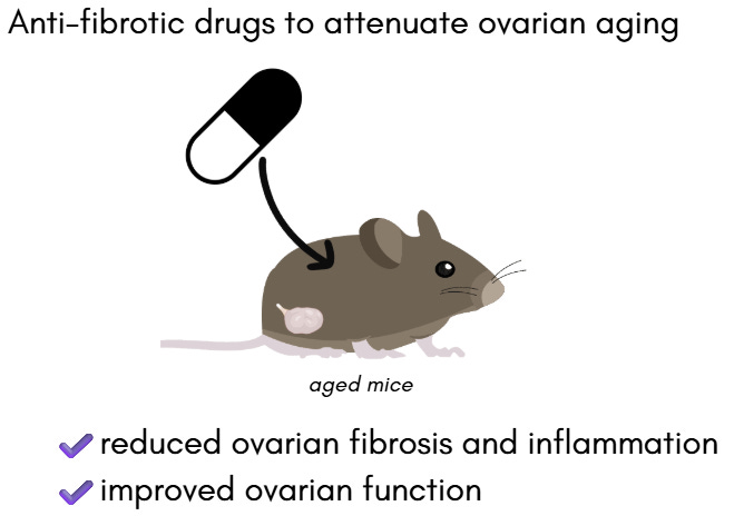
Anti-fibrotic drugs to attenuate ovarian aging
In a recent study, researchers gave aged mice a low dose anti-fibrotic drug systemically for 6 weeks. They found that compared to aged mice without the drug, there was reduced ovarian fibrosis and inflammation and improved ovarian function. This opens up the possibility of exploring drugs targeting fibrosis and ovarian stiffness to treat reproductive conditions.

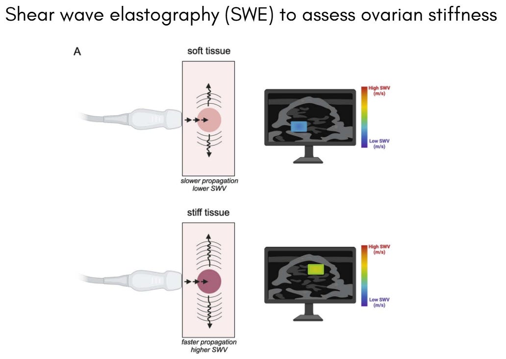
Shear wave elastography (SWE) to assess ovarian stiffness
Researchers have also proposed applying a specialized ultrasound imaging technique in a new way, to measure ovarian changes associated with reproductive aging. The ultrasound imaging technique is called shear wave elastography (SWE) which measures tissue stiffness. SWE has been used clinically as a non-invasive diagnostic tool for liver fibrosis and other pathologies. The idea would be to use SWE to non-invasively track and measure ovarian stiffness with age. Eventually, ovarian stiffness may serve as a clinical biomarker of ovarian aging which patients could use to inform their reproductive health.

Next time…
Comment below what topics you would be interested to learn more about!
If you want notifications on new posts please subscribe - it’s free! Free subscribers will also receive a visual infographic they can download about this week’s topic.
References
Schematics and illustrations created with BioRender.com and Canva
Information on inflammation in the aging ovary: https://pubmed.ncbi.nlm.nih.gov/33079460/ and https://pubmed.ncbi.nlm.nih.gov/27491879/
Repro relevance mouse study
Repro relevance shear wave elastography in ovaries paper


Share this post